Scientific Program

Conference Series Ltd invites all the participants across the globe to attend World Neuron Congress Helsinki, Finland.

Day 2 :

Keynote Forum

Ramel A Carlos

The Neurology Clinic, USA

Keynote: Obstructive sleep apnea in various cognitive disorders

Time : 10.00-11.00

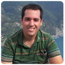
Conference Series Neuron 2018 International Conference Keynote Speaker Ramel A Carlos photo
Biography:

Ramel A Carlos is a board certified neurologist working on the Island of Guam for the past 16 years. He is currently working at The Neurology Clinic in Tamuning, Guam, USA. He completed his residency and fellowship training in Wake Forest University, Winston-Salem, North Carolina, USA. He has presented his clinical research in various neurology conferences including the Asean Neuroscience Conference in Singapore, World Congress of Neurology in London, U.K, Int. Conf. on Vascular dementia in Amsterdam, Int. Conf. of Alzheimer disease in Kyoto, Japan, and recently during the Advances in AD and PD Therapies in Torino, Italy.

Abstract:

Various research studies suggested an association between Obstructive Sleep Apnea (OSA) and various cognitive disorders,
including Alzheimer disease. The degree of OSA has been directly correlated with the severity of cognitive impairment.
Stroke and vascular diseases are significant comorbidities in these patients. We report the occurrence of OSA in patients with
various cognitive disorders on the Island of Guam and correlate the severity of OSA with the results of the neuropsychological
testing and neuroimaging studies. A retrospective review of medical records of patients evaluated in The Neurology Clinic with
the diagnosis of OSA in patients with various cognitive impairments from July 2016 to July 2018 was conducted. These include
patients with Alzheimer disease, vascular dementia, unspecified dementia, and Mild Cognitive Impairment. There were 375
patients with various cognitive impairments and 16% have been diagnosed with OSA. Among patients with OSA, 46% have
severe OSA, 38% have moderate OSA, and 16% have mild OSA. Severe impairment on Global Cognitive Scores (GCS) was seen in 60% of patients with severe OSA, 44% of moderate OSA, and 20% of mild OSA. Moderate GCS were seen in 29% of patients with severe OSA, 39% of patients with moderate OSA and 30% of patients with mild OSA. Evidences of silent stroke were seen in 25% of patients and another 31% have leukoaraiosis on their neuroimaging studies. The occurrences of vascular diseases including hypertension, diabetes mellitus, hyperlipidemia and cardiac disorders were higher in those with severe and moderate OSA compared to those with mild OSA and without OSA. Obstructive Sleep Apnea is a common comorbidity of patients with various forms of cognitive impairment. The severity of OSA correlates with the degree of impairment on neuropsychiatric testing. Neuroimaging studies demonstrated evidences of Silent stroke and leukoaraiosis among these patients.

Break: Networking and Refreshments Break 11:00-11:30 @ Foyer

Keynote Forum

Kumaar Bagrodiya

Neuroleap, India

Keynote: Using brain computer interface for improved neural activity

Time : 11.30-12.30

Conference Series Neuron 2018 International Conference Keynote Speaker Kumaar Bagrodiya photo
Biography:

Kumaar Bagrodia holds an MBA degree from the University of Oxford, is a Country Champion and Ambassador for the Oxford Business Alumni in India. He has been featured in various media in India and abroad including Knowledge at Wharton, Wall Street Journal, The Economic Times, Hindustan Times, Bloomberg TV and Forbes to name a few. He co-authored an article in Harvard HBR. He is a speaker at leading podiums including the Bombay Stock Exchange (India’s main stock exchange), has chaired international leader sessions in Israel, China Singapore and has spoken at fora with the banner of Australian Government, United Nations Environment Program, the Government of India to name a few. He was a speaker at G20 Summit yea in China 2016 and in the G20 Summit yea in Germany 2017. Kumaar was invited by the Hon’ble President of India (Pranab Mukherjee) to the official residence of the President. He was again invited in March 2018 to speak on Innovation, education and skills. He was a speaker at the Commonwealth Heads of Government Meeting in London in 2018 and was also an invitee to the palace by Royal Highness Prince of Wales.

Abstract:

TBA

  • Neurology | Neuroimaging | Neurosurgery | Neurophysiology | Neurochemistry | Neuropsychiatry and Mental Health | Neurological Disorders | Developmental Neurology
Location: Conference Hall
Speaker

Chair

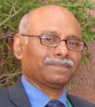
Sarwar Jamil Siddiqui

The Aga Khan University, Pakistan

Biography:

Mohammad Ghadirivasfi was graduated in psychiatry from Iran University of Medical Sciences (IUMS), Iran. He was the head of Iran Mental Hospital for 13 years and achieved years of experience in research, teaching and administration in hospital and during this period, his effort was highly effective to establish Iranian DNA Bank for Genetic and Epigenetic Studies in Psychiatric Disorders. He is interested in improving education of medical student and residency in psychiatry. He has academic publications and is one of the authors of the Iranian curriculum of general psychiatry, addiction and risky behavior fellowship and the sleep textbook (in Persian) sponsored by IUMS (Iran University of Medical Sciences). He was the secretary of 5th Basic and clinical Neuroscience Congress in 2016 Tehran, Iran.

Abstract:

rTMS, a non-invasive technique for stimulation of neuronal cells, is one of the most promising tools for brain stimulation
and modulation and reversibly affect brain cortical activity and plasticity. Although rTMS has been used to treat a variety of
neuropsychiatric disorders, several lines of evidence suggest that rTMS has a potential efficacy to reduce cue-induced craving
in drug addiction. In this study, we employed real and sham rTMS of the Orbitofrontal cortex (OFC) and the left dorsolateral
prefrontal cortex (DLPFC) to compare and test if it could reduce cue-induced craving for methamphetamine (MA) after
the end of intervention and six month follow up. Thirty eligible MA-addicted patients were recruited to receive 10 sessions
of 20min sham or 10Hz rTMS to the left OFC and DLPFC. Subjects rated their craving at baseline, after exposed to MAassociated
cues and after rTMS sessions. They were followed after six months for the rate of relapse and adherence. Relative
decrease in severity of depression was greater in OFC group, however Kruskal-Walis test showed non-significant differences
among groups (p=0.097). Also, relative changes in psychopathology severity according to the Brief Psychiatric Rating Scale,
and relative changes in craving were not statistically different among groups (p=0.350 and p=0.905, respectively) This study
failed to show any efficacy of dorsolateral or orbitofrontal rTMS in comparison with sham rTMS in reducing psychopathology
or craving of the methamphetamine dependent patients.

Biography:

Nataliia O Melnyk - the Professor of the Histology and Embryology Department of National O.O.Bogomolets Medical University, the Main Scientist in the Institute
of Genetic and Regenerative Medicine National Academy of Medical Sciences of Ukraine, Kyiv. Graduated from Kyiv National Taras Shevchenko University in
1993, after an assignment she worked as an engineer in the Institute of Molecular Biology and Genetics. During 2008- 2011, she worked as Deputy Head of the
Department of Education and Methodology of the National O.O. Bogomolets Medical University. She has more than 340 scientific and methodological works, 5
patents of scientific research. Nataliia Melnyk was edited base textbook of Histology, Cytology and Embryology for students of medical universities in Ukraine.

Abstract:

In experimental work was investigating morphological changers of neurons in organs of central nervous system (CNS) in
different experimental models of demyelination and remyelination. After induction of demyelination - EAE (experimental
allergic encephalomyelitis) - in rats, was investigate of changers of neurons in cortex of cerebrum, cerebellum and spinal
cord on 21 days and 39 days. We observed the percentage of neurons with unmodified, moderate and severe structural
changes after staining of histological sections of the brain and spinal cord by toluidine blue and cresyl violet. We studied of
demyelination process of nervous fibers in organs of CNS by the methods of electron microscopy and morphometry. We
observed of remyelination process - the percentage of normal neurons in the brain and spinal cord was increased, the amounts
of neurons with severe and destructive changes were reduce after influence of Rebif® (interferon beta-1a) by 1 and 2 weeks, and
myelinated nerve fibers was regenerate. After induction of demyelination - cuprizone model - in the 129/Sv mice, at 3-5 and 16-
17 months of age we observed changers in structure of neurons and nerve fibers. Cuprizone was provide daily for 3 weeks, in
result, was form demyelination process in CNS. RhLIF was inject after 7-days cuprizone diet, one administration daily, 50 μg/
kg, the hormone of melatonin was provide daily. In the cuprizone-treated mice of both age groups, the percentage of neurons
with severe changes in the brain and spinal cord was increased and after rhLIF and melatonin, the amounts of neurons with
destructive changes were reduce, was less pronounce in aged mice. RhLIF and melatonin may be a perspective neuroprotective drugs.

Break: Lunch break 12.30-13.30 @ Restaurant
Biography:

Sarwar Jamil Siddiqui is graduated from Dow Medical College, Karachi, Pakistan in 1985. He trained in General Medicine and Neurology at St Margaret’s Hospital,
Epping, Princess Alexandra Hospital, Harlow and St Bartholomew’s Hospital, London, England. He is also a member of American Academy of Neurology. He is
Dean, Faculty of Neurology at the College of Physicians & Surgeons of Pakistan and is responsible for Neurology Postgraduate Training in the whole country which
includes 24 training centers. This is his 4th consecutive tenure as Dean. He is also Chairman of the Editorial Board of the Journal of Pakistan Medical Association
His interests include General Neurology, Movement Disorder and Parkinson’s disease and Neurology Education and Training.

Abstract:

Pakistan is a developing country in Southeast Asia with a population of 210 million. There are about 210 practicing neurologist
in the country at present. Neurology as a specialty in Pakistan started in 1963. To start off with there were 5 neurologists,
all British trained from 1963-1969. Then more neurologists returned from abroad, especially after 1992, mainly trained in the
UK and USA. Intrinsic neurology training program started in 1990 by the College of Physicians & Surgeons, Pakistan. The first
Pakistani trained neurologist qualified in 1995. Since then the College has trained 202 neurologists to date who qualified FCPS
(Neurology) Examination after a 5-year structured training program. About 45 specialists trained in Pakistan are working
in the Middle East, UK and Canada. At present there are 21 approved neurology training centers and 24 approved training
supervisors in the country where over 120 postgraduates are undergoing training. The training includes 2 years in Internal
Medicine followed by a 3-year specialty training in Neurology. In addition to above, universities offer postgraduates programs
in Neurology namely, Diploma in Clinical Neurology (one year program) and MD (Neurology) which is a 3-year training
program in Clinical Neurology. There is a sizeable research contribution in the field of Neurology from Pakistan. The number
of research papers published in peer-reviewed journals from Pakistan was over 140 in 2017 alone.

Speaker
Biography:

Shlomi Hanassy is the owner and establisher of Hanassy R&D Ltd. IL. Shlomi is an algorithm developer and neurobiologist specialized in motor control and vision processing. He is also an inventor and experienced developer of several innovations in those fields. In 2011-2012 he worked as algorithm developer at Wellsens tech (http://www.wellsense-tech.com/), developing algorithms for pressure mat while serving as a consultant for the EU “Stiff-flop” project (http://www.stiff-flop.eu/). During 2007-2010 he was a Phd student and algorithm developer of visual substitution devises at Amir Amedi’s lab for higher brain functions while serving as a coworker in Benny Hochner’s lab for motor control and at the EU “Octopus project”(http://www.octopusproject.eu/). Since 2007 Shlomi is holding an M.sc in medical neurobiology from the Hebrew University (Hadassah Ein Carem medical school) and a B.A in computer science and administration from the Open University of Israel (since 2003).

Abstract:

Octopuses where always a fascinating animals with their spectacular line of
unique features. Here will review many of those features focusing on the
arm motor control. Similar to tongue and the elephant trunk, the octopus arm
is a muscle hydrostat, hyper-redundant, structure. The computational process
involved in the generation of a movement in such structure is a highlight
for motor control engineers. During that process, a reduction of the motor
control problem complexity is obtained by reducing degrees of freedom,
using stereotypical motor primitives, which are simply modulated instead of
calculated, to achieve the required movement. That principle inspired many
robotic engineers to achieve new kind of control: embedded intelligence. Here
we will present some results of such bio-robotic engineering and novel applications.

Break: 15.30-16.00 Networking and Refreshments Break @ Foyer
Biography:

Diana G Rotaru is currently a PhD student in the Neuroimaging Department at Centre for Neuroimaging Sciences, Institute of Psychiatry, Psychology and
Neuroscience, King’s College London. She completed her BSc at the Faculty of Medical Engineering, University Politehnica of Bucharest with a thesis focused
on EEG seizure detection using artificial intelligence. Later, her research interests turned to neuroimaging and she graduated with an MSc in Neuroimaging. She
decided to continue her dissertation project and she is working now on Magnetic Resonance Spectroscopy method development aiming to provide the tools
necessary for precise and direct measurement of low concentration neuro-metabolites on clinical and preclinical moderate-to-high-field MRI scanners.

Abstract:

As the non-invasive evaluation of brain metabolism is considered an important factor in the understanding of neurological
and psychiatric disorders (Alzheimer’s Disease, Parkinson’s Disease, epilepsy, schizophrenia, autism, ADHD, psychosis,
bipolar disorder, major depressive disorder, etc.) more interest has been given to Magnetic Resonance Spectroscopy (MRS).
Based on recent work on spectral editing schemes, discrimination and quantification of otherwise unresolvable neurometabolites
like GABA (Gamma-Amino butyric Acid, the main inhibitory neurotransmitter) and GSH (glutathione, an
antioxidant found throughout the brain) becomes possible. The purpose of our study was to test if HERMES (Hadamard
Encoding and Reconstruction of MEGA-Edited Spectroscopy) could be used to discriminate between GABA and GSH. A
comparison with standard methods (MEGA-PRESS - MEshcher-GArwood Point REsolved Spectroscopy) and test-retest
reproducibility were also considered. A special proton MRS (1H-MRS) acquisition protocol was implemented on our 3T
MR750 General Electric scanner. The HERMES pulse sequence was then tested in phantom and in vivo experiments, for the
latter, a voxel in the dorso-medial prefrontal cortex (DMPFC) being prescribed. For the measurement of the edited signal,
two software packages were used: LC Model, along with FID-A pre- processing and Gannet, which currently can be used
without any additional pre-processing steps. The acquisition results showed that spectral editing was successfully performed,
with GABA and GSH being edited correctly considering the spectral appearance and the concentration values. Regarding
the analysis methods, LC Model and Gannet were both suitable for HERMES data sets. The test-retest reliability was assessed
based on the concentration values we obtained and suggests there was a strong consistency between test-retest results for
the same method of analysis. The advantages of the HERMES pulse sequence over conventional MR protocols include time
efficiency, high-quality results and cost- effectiveness. As such, with HERMES we were able to obtain GABA and GSH spectra
simultaneously in one scanning session no longer than 12 minutes, while the time required when using MEGAPRESS for the
both GABA&GSH is double, one session of 12 minutes being necessary for each metabolite. The long-term benefits of using
HERMES include the development of a reliable tool for neuroimaging research along with improved healthcare solutions
(patient stratification, early disease detection, accurate diagnosis, treatment tracking and drug therapy evaluation).

Biography:

Rahul Srinivasan is pre final resident in Neurosurgery department at Medical Trust Hospital Kochi, Kerala. He is also member of Cochin neurological society and
neurological society of India. He has co authored various articles in field of neurotrauma. Last article related to ICP monitoring was published in Acta Neurochirugica
in 2018. During his post graduation he has assisted multiple cranial, spinal surgeries and endovascular procedure. He has won best poster award at PULSUS
Conference at Singapore conducted in September 2018. His current research also continues in field of Neuro trauma.

Abstract:

Perimedullary AV fistula is not common disease. Here we present a case of 11-year-old boy who presented with sudden
onset severe headache and neck pain for 1 day. He was having similar episodes in the past and treated as migraine. Imaging
revealed SAH in posterior fossa and cervical subarachnoid space. There were multiple flow voids around lower cervical cord
suggesting vascular malformation. Patient underwent spinal and brain DSA, which revealed diagnosis of single hole single
feeder cervical perimedullary AVF with feeding artery aneurysm as a cause of SAH. He was treated by endovascular means
with 5 month imaging follow up.

Break: Panel Discussion
  • Molecular and Cellular Neurology | Behavioural Neurology | Pediatric Neurology | Neuroendocrinology | Computational Neurology | Neuroinformatics | Neuroopthalmology
Location: Conference Hall

Session Introduction

Shlomi Hanassy

Hanassy R&D Ltd, Israel

Title: A model for the dynamics of stabilizing biological systems
Biography:

Shlomi Hanassy is the owner and establisher of Hanassy R&D Ltd. IL. Shlomi is an algorithm developer and neurobiologist specialized in motor control and vision processing. He is also an inventor and experienced developer of several innovations in those fields. In 2011-2012 he worked as algorithm developer at Wellsens tech (http://www.wellsense-tech.com/), developing algorithms for pressure mat while serving as a consultant for the EU “Stiff-flop” project (http://www.stiff-flop.eu/). During 2007-2010 he was a PhD student and algorithm developer of visual substitution devises at Amir Amedi’s lab for higher brain functions while serving as a coworker in Benny Hochner’s lab for motor control and at the EU “Octopus project”(http://www.octopusproject.eu/). Since 2007 Shlomi is holding an M.sc in medical neurobiology from the Hebrew University (Hadassah Ein Carem medical school) and a B.A in computer science and administration from the Open University of
Israel (since 2003).

Abstract:

A novel biologically inspired theory for brain information processing and encoding is presented for the first time. In this model, the world is described as networks of stabilizers components which claimed to control all biological structures, embedded in and implemented by organic systems of any form. Significant evidence for the theory is first presented in a review, and then the theory is described in an abstract way and how it is connected to information theory and qualia. And finally, preliminary results of using a simulation of the model as unsupervised ML system to solve a task are presented with comparison to classical deep learning algorithms.

Mohammad Ghadirivasfi

Iran University of Medical Sciences, Iran

Title: Neurocognitive reward circuitry system in addiction
Biography:

Mohammad Ghadirivasfi was graduated in psychiatry from Iran University of Medical Sciences (IUMS), Iran. He was the head of Iran Mental Hospital for 13 years and achieved years of experience in research, teaching and administration in hospital and during this period, his effort was highly effective to establish Iranian DNA Bank for Genetic and Epigenetic Studies in Psychiatric Disorders. He is interested in improving education of medical student and residency in psychiatry. He has academic publications and is one of the authors of the Iranian curriculum of general psychiatry, addiction and risky behavior fellowship and the sleep textbook (in Persian) sponsored by IUMS (Iran University of Medical Sciences). He was the secretary of 5th Basic and clinical Neuroscience Congress in 2016 Tehran, Iran. Currently, he is the head of neurocognitive center in Iran Mental Hospital.

Abstract:

Addiction is a chronic, relapsing disorder in which addict people mostly go through sustained habit‐based drug use and periods of abstinent. It is supposed to externalizing psychopathologies including disruptive disorders and antisocial personality disorder precedes or co-occurs with substance use disorder in at least noticeable percentage of them. Interestingly, all externalizing psychopathologies are observed from adolescence which is accompanied by risky behaviors like substance use. It is believed that three fundamental structure of neurocognitive function in children which are impaired, make them predispose to take risky behaviors and drug use. Impulsivity, decision making and working memory impairment taken together, play the main role in future brain’s reward neurocircuitry changes. Under limited inhibitory control in one hand and an overactive reward system on other hand, prone adolescences try and sustain using substance. Due to repeated intense drug rewards and neuroplasticity occurred in reward system, a compensatory anti-reward process reduces pleasurable feeling and at neuroanatomical view, a neurocircuitry migration from ventral to dorsal striatum happens. Disregarding its adverse effects and consequences, a significant percentage of addict people keep using substance. It is suggested to screen risky behavior characteristics in childhood and early adolescent in order to detect and invest on pharmacological and neurocognitive rehabilitation as a preventive program. Considering this sort of intervention from early life, it is supposed that reward system should keep unchanged until early adulthood which inhibitory system develops.

Break: Lunch Break 13.00-14.00 @ Restaurant

Mihai Stelian Moreanu & Marina Cozma

University of Medicine and Pharmacy “Carol Davila”, Romania

Title: Establishing the proper approach to an effective surgical treatment for the Meningioma
Biography:

Mihai Stelian Moreanu is currently one of the top medical students at Carol Davila University of Medicine and Pharmacy. His interests lie in neurological implications
of diseases and neurosurgery, having already completed his medical summer stage at Bagdasar-Arseni Clinical Emergency Hospital, the best neurosurgical public
center in Bucharest. His medical background includes the participation to numerous national and international congresses in Romania (IMSCB, ZEM, MEDICS) and
also abroad (FEBS 2018) gaining a vast understanding of the mechanisms of brain functioning. Also, during his medical practice he assisted to numerous surgeries
and participated to Romanian Student Surgical Society activities getting hands-on experience during surgical workshops.
 
Marina Cozma is currently studying at the Carol Davila University of Medicine and Pharmacy, being a medicine lover and an aviation enthusiast, dividing her
time between medicine and flying. She is profoundly interested in surgery and the nervous system and has just completed her summer stage at the Washington
Polyclinic in Bucharest, at the Cardiovascular Centre. Together with participating in the hardest surgeries performed in this hospital, she has perfected her
surgical skills through participating not only at national medical congresses (IMSCB, ZEM, National Anatomy Congress), but also international (FEBS 2018).
Her poster at the National Anatomy Congress was ranked in the Top Ten and she is in the process of becoming a trainer at the Romanian Student Society of Surgery.


Abstract:

Meningiomas are benign extra-axial tumors, originating from the meningeal arachnoidal cells, making up 20% of the intracranial
primary tumors. Surgical management of meningiomas is one of the most challenging procedures posing a high risk of affecting
the critical neurovascular centers of the brain. This paper attempts to identify the way Para clinical brain investigations coupled with a
well-established surgical procedure lead to an efficient and strategic treatment of meningioma, starting with a real case of a 50-year-old
woman. The clinical background of the patient was represented by frontal headaches, rare epileptic crisis, sudden dizziness, blurry vision,
loss of equilibrium and exhaustion. The Para clinical investigation included contrast MRI showing a homogenous irregular expanding
tumor process in the frontal-orbital left space. Localization, depth, diameters were also shown on MRI. Moreover, Digital Angiography
was used to identify the source of vascularization, highlighting a slight displacement of the arterial irrigation of Willis Polygon towards
the tumoral process. These symptoms and investigations led to diagnosing a sphenoid left wing meningioma. The treatment was mainly
focused on the neurosurgical intervention, having several purposes: rejecting the meningioma, establishing the anatomo pathological
diagnosis, developing the therapeutic plan. The surgical procedure included the following steps: positioning, incision, craniotomy,
tumor exposure, devascularization, decompression, extra capsular dissection, tumor removal, closure. The surgical approach was on
the frontotemporal side and after a step-by-step incision, the tumor was exposed. The excision strategy was focused on fragmentation
and mild dissection of the surrounding brain tissue. Also, while the tumor surface coagulated constantly, it lost volume and allowed an
easy dissection. The final result was favorable – the patient regained her balance. Getting a better understanding of the neurosurgical
steps of treating meningiomas will lead to finding strategies that will improve the patient's treatment and his quality of life.

Break: 15.00-15.30 Networking and Refreshments Break @ Foyer